
Description
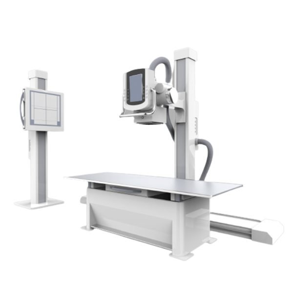
Double-Column DR Machine Feature
Double column design with ground rail, large range of projection area adjustment space, easily meet the various shooting needs
Intelligent operation, lets you experience the ease and fun of work; Automatic optimization of tube tracking software system, and organization of the balanced processing technology, bring you a brand new experience
1. Flat Panel Detector
Type of the detector: TFT monolithic amorphous silicon
Panel size of the detector: 430mm*430mm
Efficient imaging size of the detector: 17*17 inch
Acquisition pixel matrix of the detector: ≥3072×3072
The pixel pitch of the detector: ≤146 um
The spatial resolution of the detector: 3.7LP/MM
Pixel grayscale: 16-bit
Time of image acquisition and transmission: 3s
2. High frequency and high voltage generator
Max. Output power: 50kw
Input power supply: 380 VAC three-phase
Output voltage: 40~150kV
Tube current range: 10~800mA
Exposure time range: 1ms-10000ms
mAs range: 0.1mAs~1000mAs
Support AEC、APR automatic exposure
Diagnostic Self-test and display
3. X-ray bulb tube assembly
Focus: 0.6mm/1.5mm
Nominal electric power: 21kVA/52kVA
Max. KV: 150kV
Anode target angle: 12°
4. Bed for radiography
The longitudinal movement range: ±130 mm
The lateral movement range: is ±280 mm
5. Vertical chest X-ray stand
Vertical movement range: 1400±20 mm
6. Radiography stand
Rotation range of the tube assembly: ≥±90°; Positioning for each 90°
Longitudinal movement range: 1640±20 mm
Vertical movement range: 1180±20 mm
7. AEC (optional)
The detection number of the outliers: 3
Tube voltage range: 40-150KV
Light time range: 1ms-10s
8. The grid and the beam-limiting device
The grid material: Aluminum-based grid
Size: 18×18 inch (48×48cm)
Grid ratio: 10:1
Density: 215C
Grid Focal length: 1000mm
9. Workstation functions for image acquisition, processing, and diagnosis
The Chinese user interface, standard DICOM3.0 image
Workstation functions for image acquisition: adjust or preset window width/ window level, local automatic window level, preset window width/ window level, positive and negative image flipping, image flipping, rotation, image magnification and roam, image interpolation edge enhancement, local magnification, restoration, image annotation, text annotation/number annotation, image marking, ruler line segment measurement, square and round measurement, arbitrary shape measurement, angle measurement, automatic electron sharing, image stitching, acquisition and display of exposure index
Software package for the special image acquisition control and software package for special parts protocol processing
Functions of patient management, image acquisition, image processing (image correction, image flipping, USM sharpening, image filtering), image observation (provide the tools for observation and measurement), image stitching
Image printing, DICOM printing, paper printing, manual printing for the displayed images, a single button for marking and printing the images, optional for different printers, film format, and number of prints, print queue control, stop/start the preset
User personalization: showing the format and layout, default setting, toolbar setting, and parts protocol enhancement filter
Image display: display configuration 1920×1080, HD display
10. Workstation
CPU: Intel i5,3GHz or above
Host memory: ≥8GB DDR4
Hard disk: 1T/7200rpm large capacity and high-speed hard disk
Workstation monitor: 24” IPS LCD monitor
Network card and network interface: 1000M network card, 1000M network interface, Jumbo Frames: 9K
DICOM3.0 interface
Windows 10 64-bit SP1 (Professional Edition or higher)
Related Products

Reviews
There are no reviews yet.